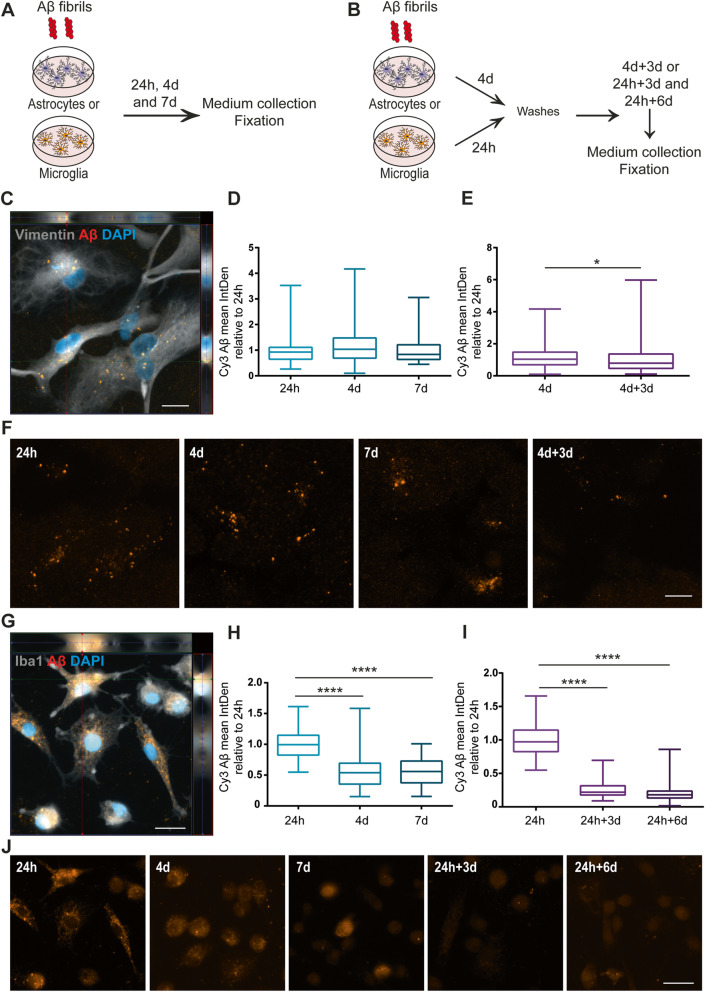
Fig. 3.
Intracellular Aβ levels are reduced in microglia over time but remain stable in astrocytes. Schematic figure of the study design illustrating that the cells were either constantly treated with Aβ-F for 24h, 4d, and 7d (a) or treated with an Aβ-F pulse (b), lasting for 24h (microglia) or 4d (astrocytes), followed by culture in Aβ-F-free medium. Astrocytes had ingested and accumulated Aβ-F already at 24h (c). The intracellular Aβ accumulation was significantly increased after 4d compared to 24h (d). At 4d+3d, the astrocytic Aβ signal was significantly reduced, compared to the 4d time point (e). Representative images of the Aβ deposits in astrocytes at the different time points are shown in f. Large amounts of intracellular Aβ could be detected in microglia at 24h (g). Contrary to the astrocytes, microglia showed a reduction in intracellular Aβ signal at 4 days and 7d compared to 24h (h). Furthermore, the intracellular Aβ aggregates in microglia were significantly lowered at 24h+3d and 24+6d, compared to 24h (i). Representative images of the Aβ deposits in microglia at the different time points are shown in j. Scale bars = 20μm

