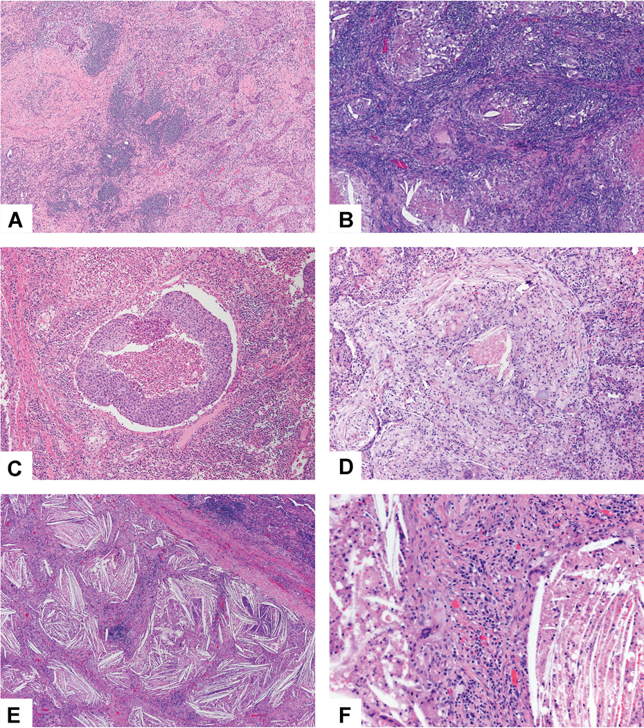
Figure 11:

Stromal inflammation and necrosis. A) This squamous cell carcinoma has numerous lymphoid aggregates in the stroma. B) This adenocarcinoma shows a dense lymphoplasmacytic stromal infiltrate. C) Numerous neutrophils are seen not only within the focus of tumor necrosis in the center of the image, but also within the surrounding stroma. D) The tumor stroma is infiltrated by numerous histocytes which show a small area of necrosis in the center. E) Prominent cholesterol clefts are seen in this area of the tumor bed. F) The cholesterol clefts are surrounded by bands of stroma with prominent chronic inflammation and giant cells, some of which are associated with the cholesterol clefts.
