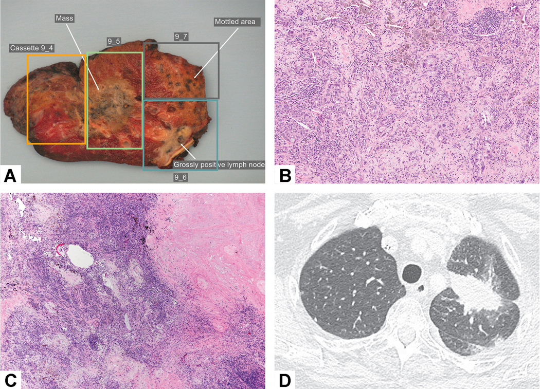
Figure 6:

Tumor bed with multiple areas of viable tumor. The viable tumor alternated with stromal inflammation and fibrosis that led the person grossing the specimen to describe three different areas of tumor (in blocks 9–4, 9–5 and 9–7) raising the question whether there were intrapulmonary metastases. An area in block 9–6 shows grossly positive lymph nodes reflecting metastatic carcinoma. B) Residual viable tumor consists of acinar glands and the adjacent stroma shows prominent chronic inflammation and loose myxoid connective tissue. C) The nodules of residual viable tumor alternated with intervening fibroinflammatory stroma in the tumor bed. D) Prechemotherapy CT scan shows a solitary mass confirming that there is a single tumor with a heterogeneous response to chemotherapy.
