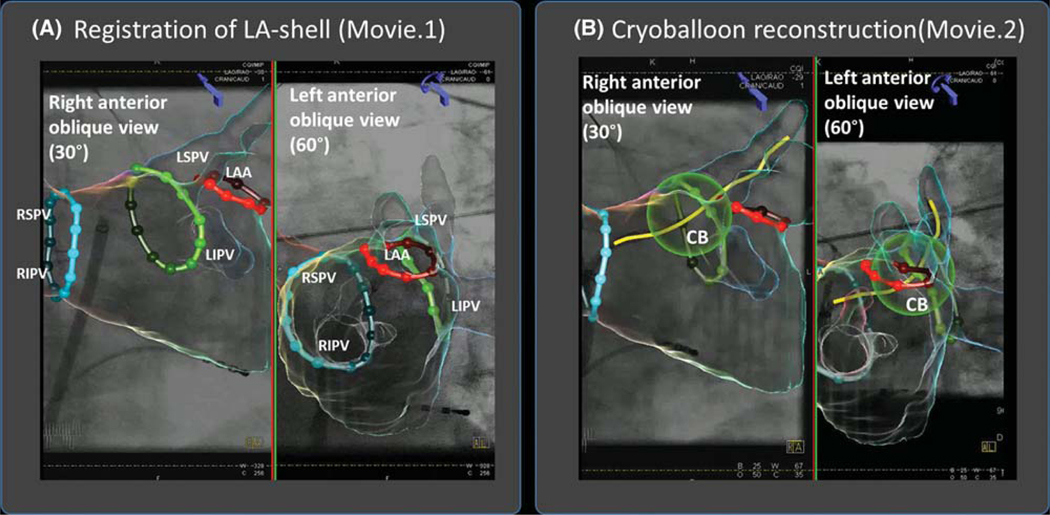
FIGURE 1.
Novel overlay guidance system superimposes left atrial anatomy on fluoroscopic images during cryoablation procedure. A, Registration of LA-shell segmented from preprocedural CT on biplane fluoroscopic view (and Movie 1) in 30◦ right anterior oblique and 60° left anterior oblique views. Contrast is injected and the model is fit to the outline of the contrast cloud in both views. B, Cryoballoon reconstruction (and Movie 2). From this step, the cryoballoon catheter is identified in both views by manually marked points (proximal and distal ends of the balloon, and the distal end of the wire), which then allow automatic balloon reconstruction Abbreviations: CB = cryoballoon; LAA = left atrial appendage; LIPV = left inferior pulmonary vein; LSPV = left superior pulmonary vein; RIPV = right inferior pulmonary vein; RSPV = right superior pulmonary vein

