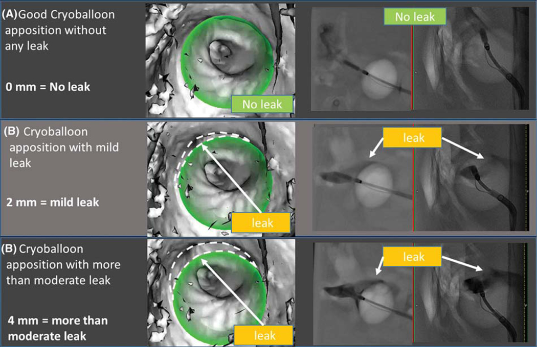
FIGURE 4.
Results of phantom experiment: Leak detection by overlay guidance system and venogram: (A) No leak; (B) mild leak (2 mm); (C) more than moderate (4 mm). The novel imaging system (column 1) shows the reconstructed cryoballoon (in green) in the left atrial shell. The imaging system accurately identified apposition (a) or leak dimensions in each case (B and C). Venography (column 2) shows biplane cineangiograms during contrast infusion, again showing identifiable leaks (B and C) and albeit with difficulty to quantify size. Dashed lines were added to the figure to depict leaks; these are not part of the original image generated by the overlay guidance system

