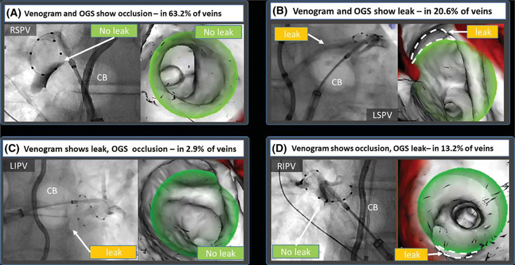
FIGURE 5.
First venogram performed after cryoballoon apposition at each vein. In the majority of cases (83.8%), there was concordance between venogram and the overlay guidance system (OGS). A, In 63.2% of PVs, both methods showed occlusion. C, The majority of leaks (20.6%) were detected by both venography and the OGS. C, In 2.9%, the OGS did not show a leak detected by venography. D, In 13.2% of PVs, the OGS was able to detect leaks missed by venogram. Dashed lines were added to the figure to depict leaks, these are not part of the original image generated by the overlay guidance system

