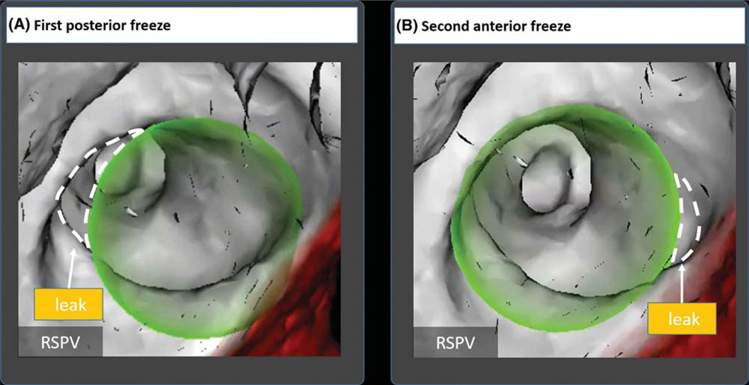
FIGURE 6.
Two-step cryoballoon isolation of right superior pulmonary vein in a 72-year old woman. A, Initial positioning showed an anterior leak by venography confirmed by the overlay guidance system (OGS). Staged cryoballoon therapy started with therapy delivery to the posterior portion of the RSPV. B, The cryoballoon was then repositioned to cover the anterior RSPV by venography and confirmed on OGS. The OGS was able to visualize elimination of selected gaps by repositioning of the cryoballoon in real time. Dashed lines were added to the figure to depict leaks; these are not part of the original image generated by the overlay guidance system

