
Figure 4.
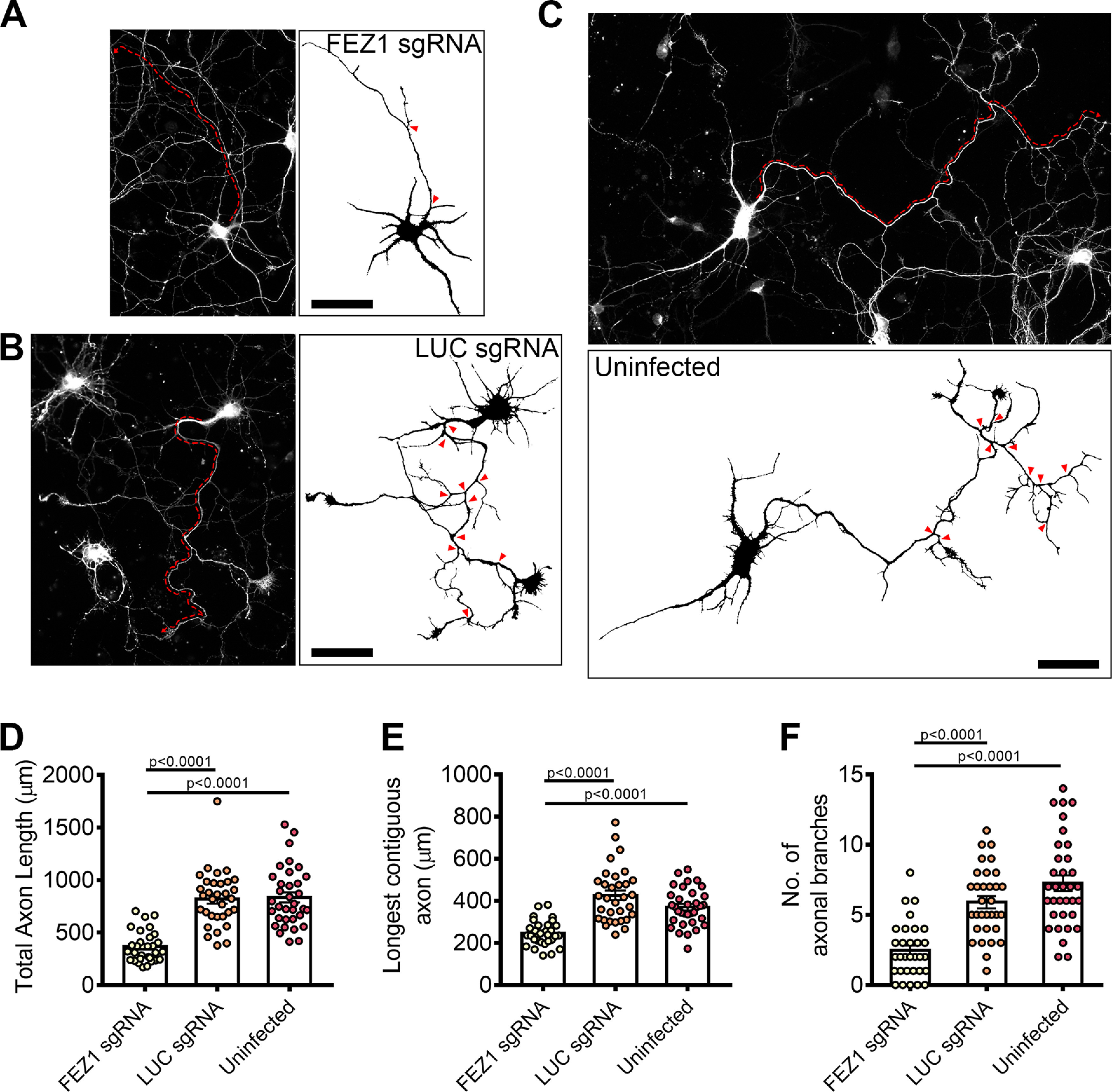
FEZ1-deficient neurons display defects in axonal development. A–C, Tiled images of FEZ1 sgRNA, LUC sgRNA, and uninfected neurons immunostained for Tau. Respective axon traces are shown juxtaposed. Axons of FEZ1-deficient neurons are significantly less branched as compared with control neurons (branch points indicated by red arrowheads). The length of the longest contiguous axon (red dotted lines) in FEZ1-deficient neurons was also shorter than in control neurons. D–F, Quantification of total axon length, longest contiguous axon length, and number of axon branches in FEZ1 sgRNA, LUC sgRNA, and uninfected neurons, respectively. All quantified variables were significantly decreased in FEZ1-deficient neurons as compared with control groups. Statistical significance was determined using Kruskal–Wallis analysis. FEZ1 sgRNA n = 31, LUC sgRNA n = 32, uninfected n = 34, obtained from three independent experiments. Error bars represent SEM. Scale bars: 50 μm. Developmental abnormalities in shRNA-mediated FEZ1 knock-down hippocampal neurons are shown in Extended Data Figure 4-1.