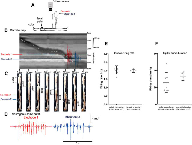
Figure 10.
Electrical recordings from colonic smooth muscle during propulsion of fecal pellets in an isolated whole mouse colon. A, Schematic of the whole colon with natural colonic content. A video camera recorded colonic wall movements and two extracellular recording electrodes recorded smooth muscle action potentials as the fecal pellets were propagated past the electrodes. B, A spatiotemporal map of fecal pellet propulsion. The red and blue arrows show the relative location of the electrodes when the pellet propagates past the recording site. C, A series of photomicrographs as single fecal pellets propagate past the two recording sites. The red and blue arrows show the location of electrical recording sites. D, The actual discharge of action potentials in the smooth muscle in red and blue that represent the activity in the muscle as the fecal pellet is propelled past the recording. Note, the action potentials discharge in rhythmic bursts at ∼2 Hz. This can also be observed in Movie 4. E, The mean action potential firing rate in tubular intact preparations of whole colon versus in flat-sheet preparations of whole colon. F, The action potential burst duration in the smooth muscle as content is propelled along intact tube preparations of colon compared with flat-sheet preparations.

