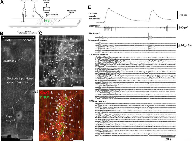
Figure 4.
Simultaneous neuronal imaging and smooth muscle electrophysiology during propagating neurogenic spike bursts in whole mouse colon. A, Schematic of the location of the neuronal imaging and electrophysiological recordings. Two extracellular recording electrodes were positioned 15 mm apart along the length of colon. B, The full circumference of mouse colon and the location of the myenteric ganglia imaged relative to the position of electrode 2. Calibration, 1 mm. C, Fluo-4 in the live preparation showing the recorded neurons and internodal strands. Calibration, 100 μm. D, shows the neurochemical coding of the region shown in C. Numerous excitatory (ChAT+ve in red) and inhibitory (NOS+ve in green) neurons are shown in neighboring myenteric ganglia. Calibration, 100 μm. E, shows periodic neurogenic contractions and propagating bursts of muscle action potentials recorded from electrodes 1 and 2. Before and during the neurogenic contractions, neuronal imaging showed increased activity in internodal strands, ChAT+ve and NOS+ve neurons. Figure 4-1 shows that synchronized firing of ChAT+ve and NOS+ve neurons was also temporally coordinated with activation of motor nerve axons in the circular muscle and occurred in all detectable nerve cell bodies.

