Figure 6.
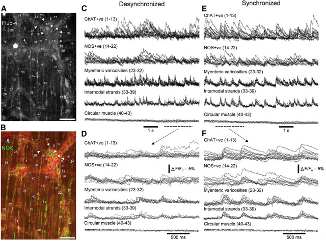
Two distinct periods of synchronized and desynchronized neuronal activity occurred in the myenteric plexus during propagating neurogenic contractions. A, The structures (dotted outlines) from which calcium recordings were taken. These include ChAT+ve nerve cell bodies (numbered 1–13), NOS+ve nerve cell bodies (14–22), myenteric varicosities (22–32), internodal strands (33–39), and sections of circular muscle (40–43). Recordings were made in the presence of nicardipine (1 μm) to paralyze the smooth muscle. B, The neurochemical coding of the imaging region shown in A. Scale bar, 100 μm. C, During the intervals between neurogenic contractions, there was weak temporal coordination of calcium transients from cholinergic and nitrergic neurons, myenteric varicosities and internodal strands. Scale bar, 100 μm. D, The period corresponding to the dashed bar in C. Relatively weak temporal coordination between the neuronal structures occurred. E, Activity from the same sources in A, but with temporal coordination between the different classes of nerve cell bodies, internodal strands, and varicosities. F, Expanded region of the recording period represented by the dotted line in E. Comparison of recordings E and F shows the changes in coordination of neuronal firing between desynchronized and synchronized states. Of particular interest was the observation that during synchronized neuronal activity, CGRP+ve myenteric neurons also became temporally activated with all the ChAT+ve and NOS+ve neurons, as shown in Figure 6-1.

