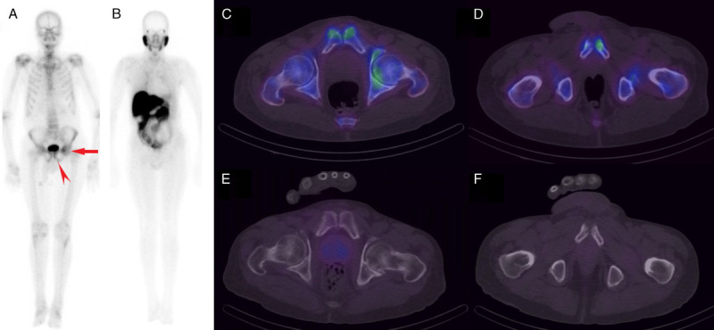
FIGURE 2.

Whole-body 99mTc-MDP scan (A) showing asymmetrically increased uptake in the left pubis (arrowhead) and iliac region (arrow), whereas no obvious increased uptake is seen in the corresponding areas on 99mTc-PSMA scan (B). SPECT/CT through 2 different slices highlighting the above changes localizes the iliac findings to the left femoroacetabular joint region (C) in keeping with degenerative change. The focal uptake seen in the left pubis (D) localizes to an area of sclerosis adjacent to the pubic symphysis. This was of concern for metastasis; however, degenerative change was also a consideration due to the proximity to the joint (rendering the finding equivocal). On 99mTc-PSMA SPECT/CT scans (E and F), there was no pathological uptake in the corresponding areas to suggest osseous metastases.
