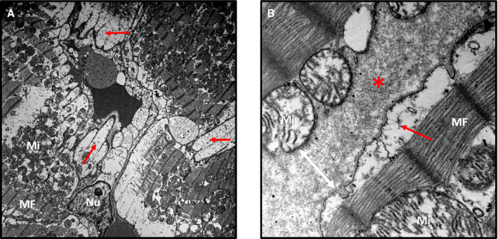
Figure 4. Interstitial and cytoplasmic edema after irradiation.

A, Rat left ventricular midmyocardium on electron microscopy (×5000 magnification). B, Rat left ventricular midmyocardium on electron microscopy (×40 000 magnification), left ventricle irradiated with 30 Gy (3 weeks). Cellular membranes were dilated because of cytoplasmic swelling (red arrows). Interstitial edema widened intercellular spaces (red asterisks and white arrow). Subsarcolemmal and interfibrillar mitochondria (marked as “Mi”) were damaged and variable in size. Myofibrils (MFs) with Z and M lines were intact. No remarkable abnormalities in myocyte nuclei (marked as “Nu”) were noted.
