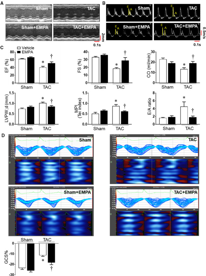
Figure 2. Empagliflozin treatment improved cardiac function and exercise endurance in heart failure.

(A) and (B) Representative images of M‐mode of left ventricles and mitral valve flow Doppler in Sham and transverse aortic constriction groups. (C) Cardiac systolic and diastolic function were quantified, including ejection fraction, fraction shortening, cardiac output, myocardial performance index, and E/A ratio. The end‐diastolic left ventricular posterior wall thickness was also evaluated. (D) Cardiac speckle‐tracking analysis was used to evaluate cardiac contractility. Upper: the representative images of global circumferential strain among groups. Lower: Quantitative results. One‐way ANOVA (non‐repeated measures) (C–D). Results are expressed as mean±SEM, n=5–6 in each group. CO indicates cardiac output; EF, ejection fraction; FS, fraction shortening; GCS, global circumferential strain; LVPWd, LV posterior wall during diastole; and TAC, transverse aortic constriction. *P<0.05 vs sham, † P<0.05 vs transverse aortic constriction.
