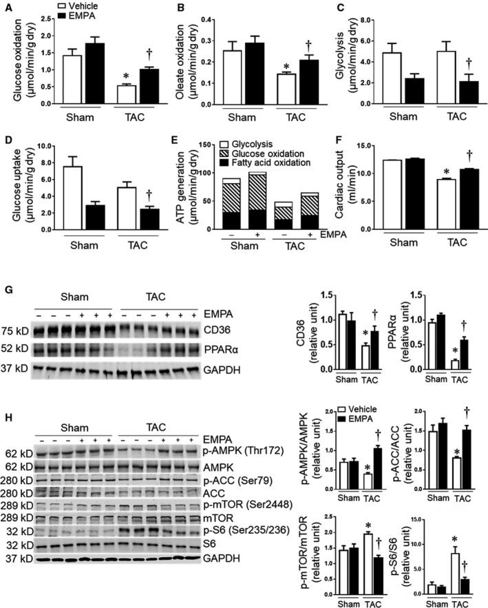
Figure 5. Chronic empagliflozin treatment improved oxidative phosphorylation in failing hearts.

(A) to (D) Mean glucose and oleate acid oxidation, glycolysis, and glucose uptake in isolated perfused hearts from mice in different groups at the end of the protocol (n=5–6). (E) Calculated ATP generation in Sham, Sham+empagliflozin, TAC and TAC+empagliflozin groups (n=5–6). (F) The cardiac output of the isolated perfused hearts (n=5–6) in each group. (G) Left: representative blots of CD36, PPARα, and GAPDH. Right: Quantitative analysis (n=5–6). (H) Left: the representative blots of AMP‐activated protein kinase, acetyl‐coA carboxylase, mammalian target of rapamycin complex 1, S6 ribosomal protein and GAPDH were shown. Right: quantitative analysis (n=5–6). Results are expressed as mean±SEM; One‐way ANOVA (non‐repeated measures) (A–H). ACC indicates acetyl‐coA carboxylase; AMPK, adenosine monophosphate‐activated protein kinase; CD36, cluster of differentiation 36; mTOR, mammalian targe of rapamycin; S6, ribosomal protein S6; and TAC, transverse aortic constriction. *P<0.05 vs sham, † P<0.05 vs transverse aortic constriction.
