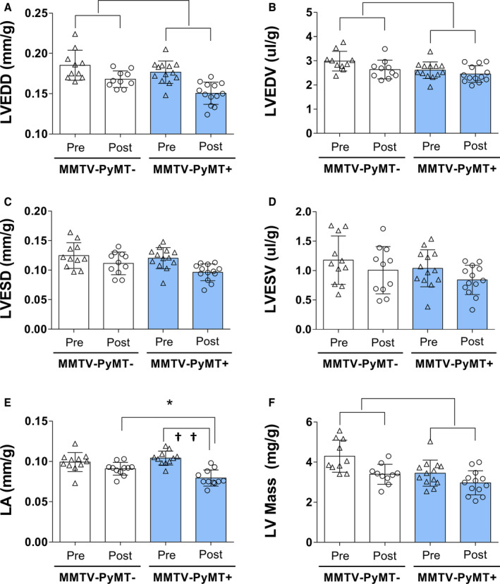
Figure 2. Morphological and functional cardiac parameters in mouse mammary tumor virus–polyomavirus middle T antigen (MMTV‐PyMT)− and MMTV‐PyMT+ mice in the pre‐experimental and post‐experimental protocol.

Left‐ventricular (LV) end‐diastolic diameter (LVEDD) (A); LV end‐diastolic volume (LVEDV) (B); LV end‐systolic diameter (LVESD) (C); LV end‐systolic volume (LVESV) (D); left atrium (LA) (E); and LV mass (F). Parameters were corrected by weight. LVEDD: P group<0.001, P interaction=0.417; LVEDV: P group=0.003, P interaction=0.437; LA: P interaction=0.004; post hoc test: * P=0.030, between group difference and †† P<0.001, within MMTV‐PyMT+ group difference; and LV mass: P group<0.001, P interaction=0.365. Generalized estimation equations with normal distribution and identity link function using AR(1) correlation matrix between evaluations followed by Bonferroni multiple comparison. Values are expressed as means and SD. Four mice in the MMTV‐PyMT− and 2 mice in the MMTV‐PyMT+ were excluded from this analysis because of poor imaging quality on echocardiography. Thus, 10 mice in the MMTV‐PyMT− and 13 mice in the MMTV‐PyMT+ were involved in this analysis. Of note, 2 LA measures were not obtained in the MMTV‐PyMT+ mice.
