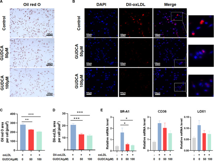
Figure 1. GUDCA inhibits macrophage‐derived foam cell formation.

THP‐1 macrophages were pretreated with indicated concentrations (0, 50, or 100 μM) of GUDCA, then incubated with oxLDL or DiI‐labeled oxLDL for 24 h. A, Representative Oil red O staining in oxLDL‐induced THP‐1 macrophages. B, Fluorescent images of THP‐1 macrophages incubated with DiI‐labeled oxLDL. Scale bar, 100 μm. C, Quantitative analyses of Oil red O and (D) DiI‐oxLDL positive area (representative as pixel2 per cell). E, Relative mRNA expression of genes involved in oxLDL uptake; GAPDH mRNA was used as internal control. Data are representative of three independent experiments and shown as mean±SEM. Statistical analyses were performed with 1‐way ANOVA and followed by Dunnett multiple comparisons. *P<0.05, **P<0.01 and ***P<0.001 vs control. DiI‐oxLDL indicates 1,10‐dioctadecyl3,3,30,30‐tetramethylindo‐carbocyanine‐labeled oxidized low‐density lipoprotein; GUDCA, glycoursodeoxycholic acid; LOX‐1, lectin‐like oxidized low‐density lipoprotein receptor‐1; oxLDL, oxidized low‐density lipoprotein; and SR‐A1, scavenger receptor A1.
