
Figure 1. Adipocyte sizes in fat pads.
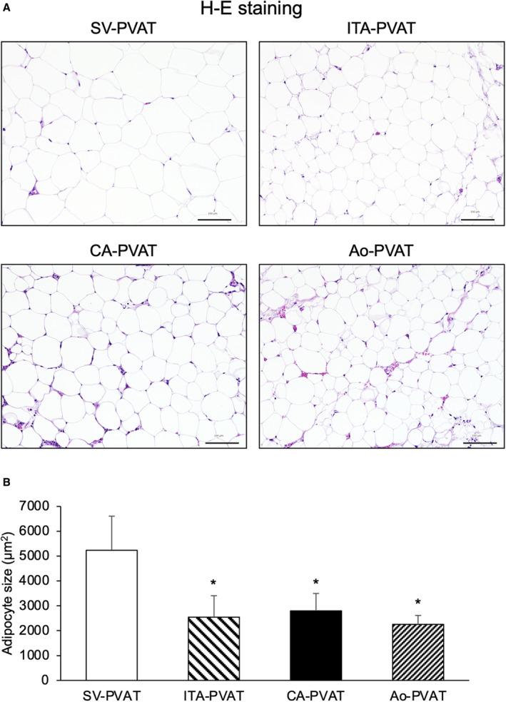
A, Representative hematoxylin‐eosin (H‐E) staining of perivascular adipose tissue surrounding the saphenous vein (SV‐PVAT), that surrounding the internal thoracic artery (ITA‐PVAT), that surrounding the coronary artery (CA‐PVAT), and that surrounding the aorta (Ao‐PVAT). Bar=100 µm. B, Comparison of adipocyte sizes in SV‐PVAT, ITA‐PVAT, CA‐PVAT, and Ao‐PVAT of patients (n=48). Results are shown as means±SD. *P<0.05 vs SV‐PVAT.