
Figure 1. CD11b is upregulated in human AAA tissues and a CaCl2‐induced AAA model.
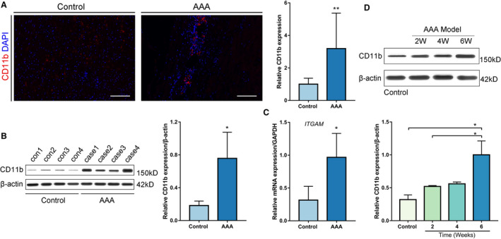
A, Representative confocal immunofluorescence staining of CD11b and DAPI in human AAA tissues and control tissues. Scale bars=100 μm. Quantification of CD11b based on the integral optical density in AAA (n=4) and control (n=4) tissues, **P<0.01 vs control. B, WB and densitometric analysis of CD11b protein expression in human AAA samples (n=4) and control samples (n=4), *P<0.05 vs control. C, Quantitative real‐time polymerase chain reaction for CD11b mRNA expression in human AAA tissues (n=4) and nonaneurysmal abdominal aortic tissues (n=4). *P<0.05 vs control. D, WB and densitometric analyses of CD11b protein expression in abdominal aortic tissues from control wild‐type mice, and from mice 2 weeks, 4 weeks, and 6 weeks after AAA establishment, *P<0.05 vs control or 2 weeks. AAA indicates abdominal aortic aneurysm; DAPI, 4',6‐diamidino‐2‐phenylindole; ITGAM, integrin subunit alpha M; and WB, Western blot.