
Figure 3. ITGAM deficiency ameliorates macrophage infiltration and polarization.
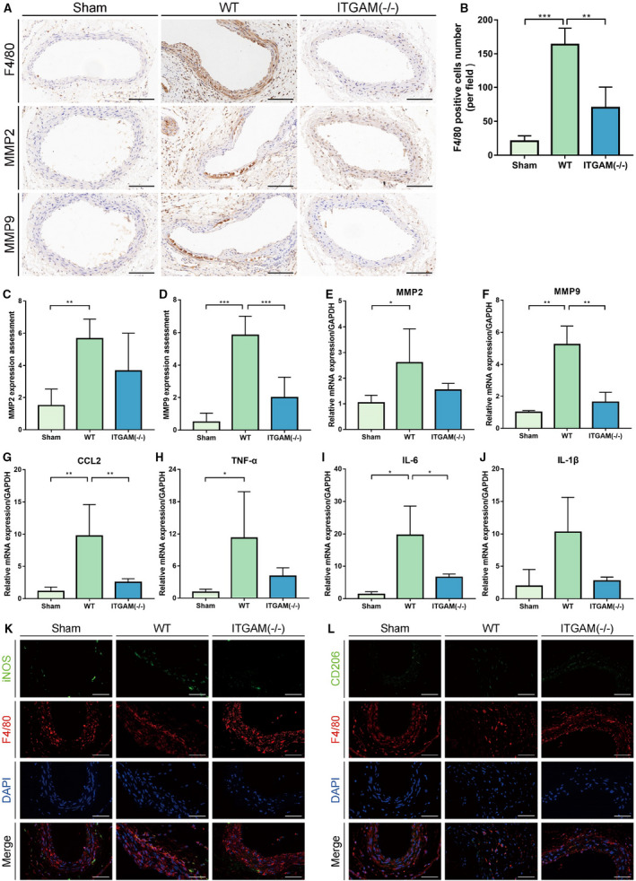
A, Representative images of immunohistochemical staining for F4/80, MMP2, and MMP9 in the aortic tissues of saline‐ or CaCl2‐treated WT or ITGAM(‐/‐) mice. Scale bar=100 μm. B, Quantification of F4/80‐positive macrophages in the infrarenal abdominal aortas of the mice from the 3 experimental groups under a microscope (n=6 for each group), **P<0.01, ***P<0.001. C and D, Quantification of MMP2 and MMP9 immunostaining in the abdominal aortic walls of the experimental mice (n=6), **P<0.01, ***P<0.001. E through J, Relative MMP2, MMP9, CCL2, TNF‐α, IL‐6, and IL‐1β mRNA expression in the abdominal aortic walls of the experimental mice (n=3), *P<0.05, **P<0.01. K and L, Representative images of dual immunofluorescence staining of iNOS (green), CD206 (green), F4/80 (red), and DAPI (blue) in the abdominal aortas of WT and ITGAM(‐/‐) mice 6 weeks after exposure to CaCl2. Scale bars=50 μm. DAPI indicates 4',6‐diamidino‐2‐phenylindole; IL‐6, interleukin 6; IL‐1β, interleukin 1β; iNOS, inducible nitric oxide synthase; ITGAM, integrin subunit alpha M; MMP, matrix metalloproteinase; TNF‐α, tumor necrosis factor α; and WT, wild‐type.