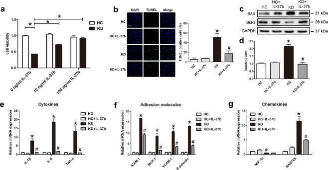
Fig. 3. IL-37b alleviated KD-treated endothelial cell apoptosis and inflammation.
a CCK8 assay was used to evaluate cell viability (n = 6). Significance: *P < 0.05. b DNA fragmentation was analyzed using TUNEL staining (n = 3). c Protein expression of BAX and Bcl-2 was determined by western blot analysis. d Quantitative analysis of BAX/Bcl-2 ratio was conducted. Data are presented as mean ± SD (n = 3). e, f The expression levels of cytokines, adhesion molecules, and chemokines were examined in KD-treated ECs after IL-37b treatment (n = 3). *P < 0.05 vs. the HC group, and #P < 0.05 vs. the KD group. At least three experiments were performed for each assay.

