
Figure 2. Spectral-Domain Optical Coherence Tomographic Images of Patients With Achromatopsia and a Representative Control Participant Aged 1.3 Years.
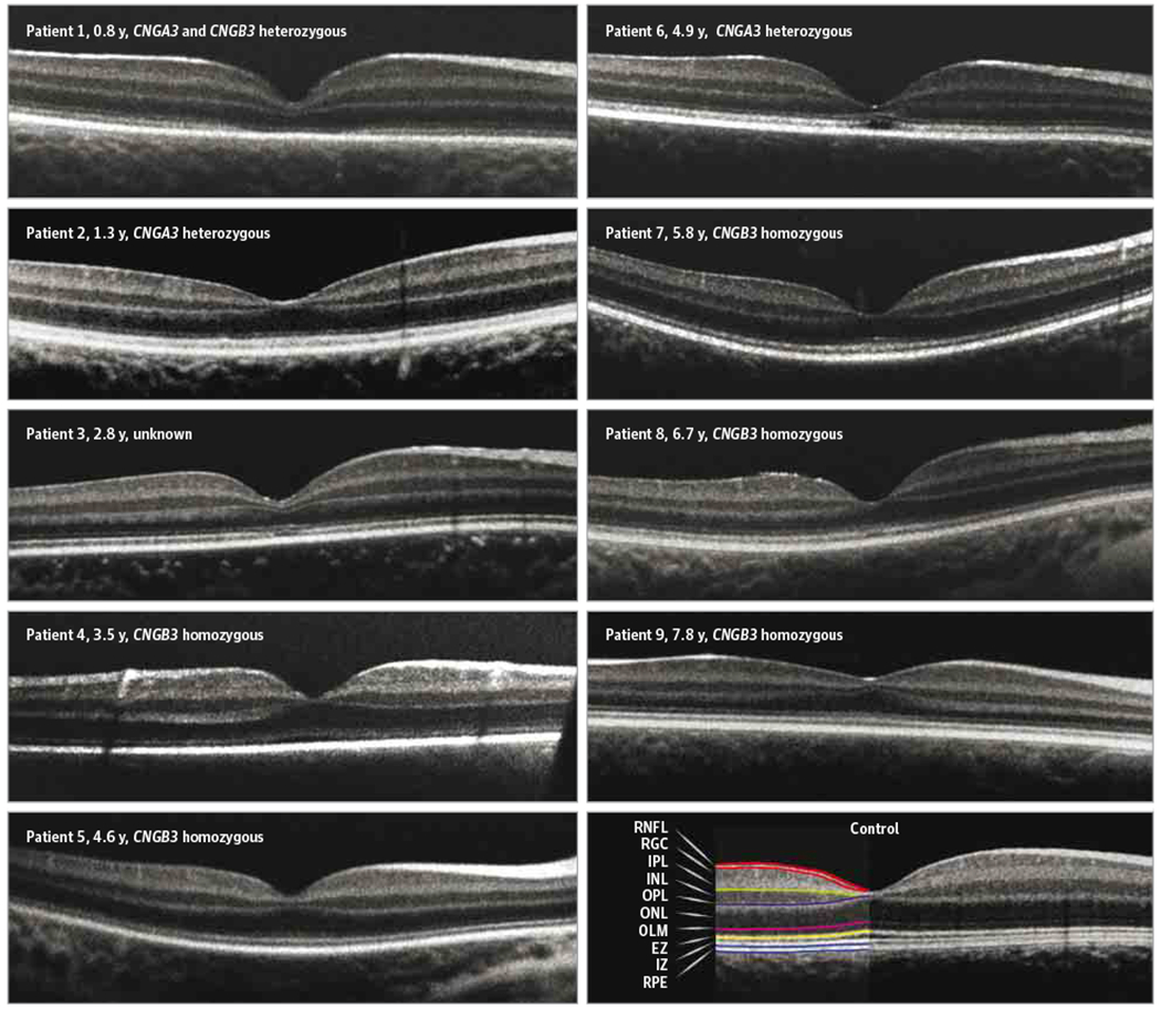
The ages and genotypes are indicated for each patient with achromatopsia. In this young cohort, axial length measurement was not possible owing to time and technical constraints during the sedation protocol, so a common scale bar was not applied. EZ indicates ellipsoid zone; INL, inner nuclear layer; IPL, inner plexiform layer; IZ, interdigitation zone; OLM, outer limiting membrane; ONL, outer nuclear layer; OPL, outer plexiform layer; RGC, retinal ganglion cell; RNFL, retinal nerve fiber layer; and RPE, retinal pigment epithelial layer.