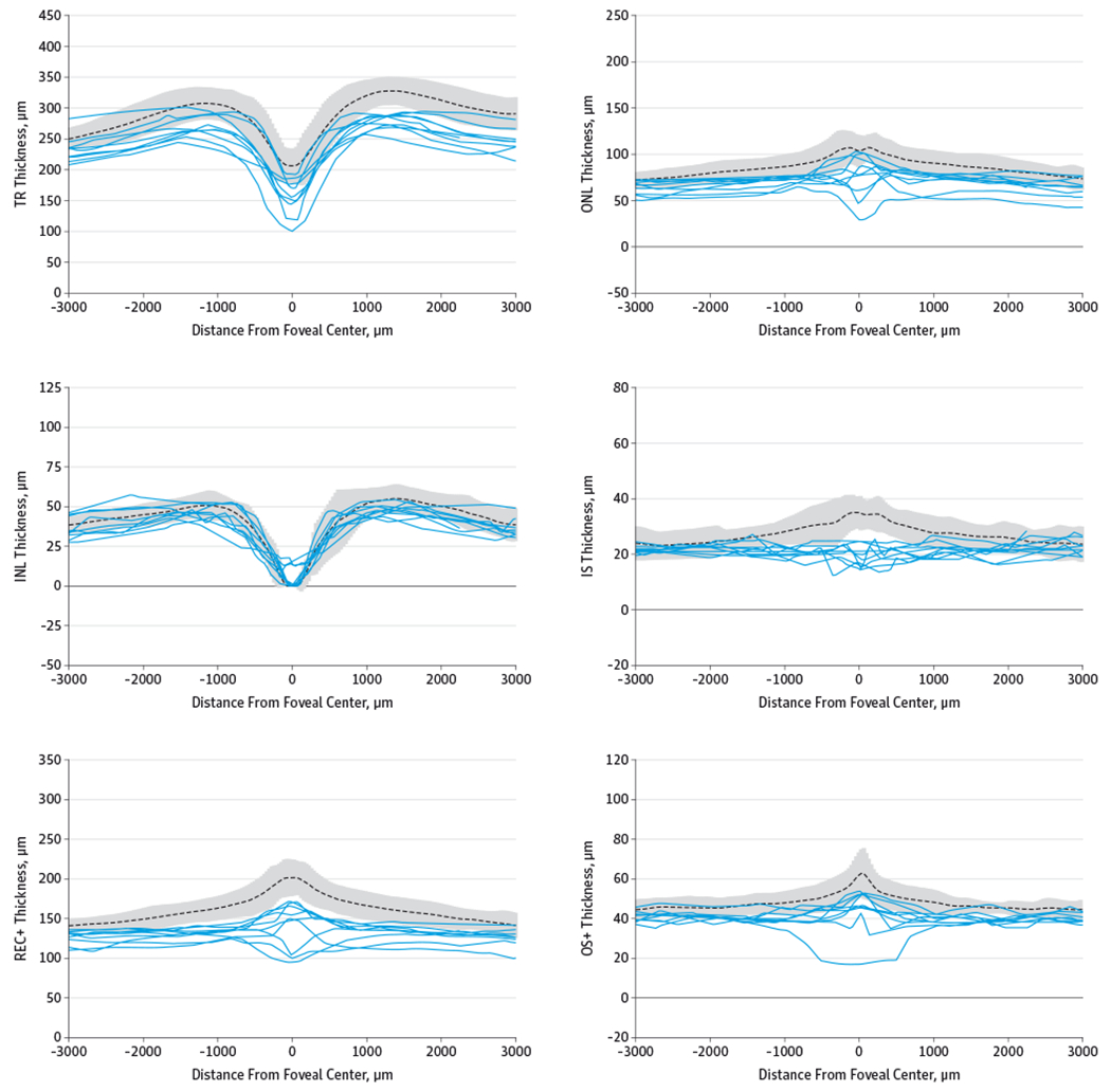
Figure 4. Retinal Layer Thicknesses of Patients With Achromatopsia Plotted Against the Control Mean ± 2 SDs.

Retinal layer thicknesses are shown for patients with achromatopsia (solid lines) plotted against the control mean (dotted line) ± 2 SDs (gray area). INL indicates inner nuclear layer; IS, inner segment layer; ONL, outer nuclear layer; OS+, outer segment plus retinal pigment epithelial layer; REC+, photoreceptor layer from Bruch membrane to INL/outer plexiform interface; and TR, total retina. For distance from foveal center, −3000 is in the direction of temporal retina and +3000 is nasal retina.
