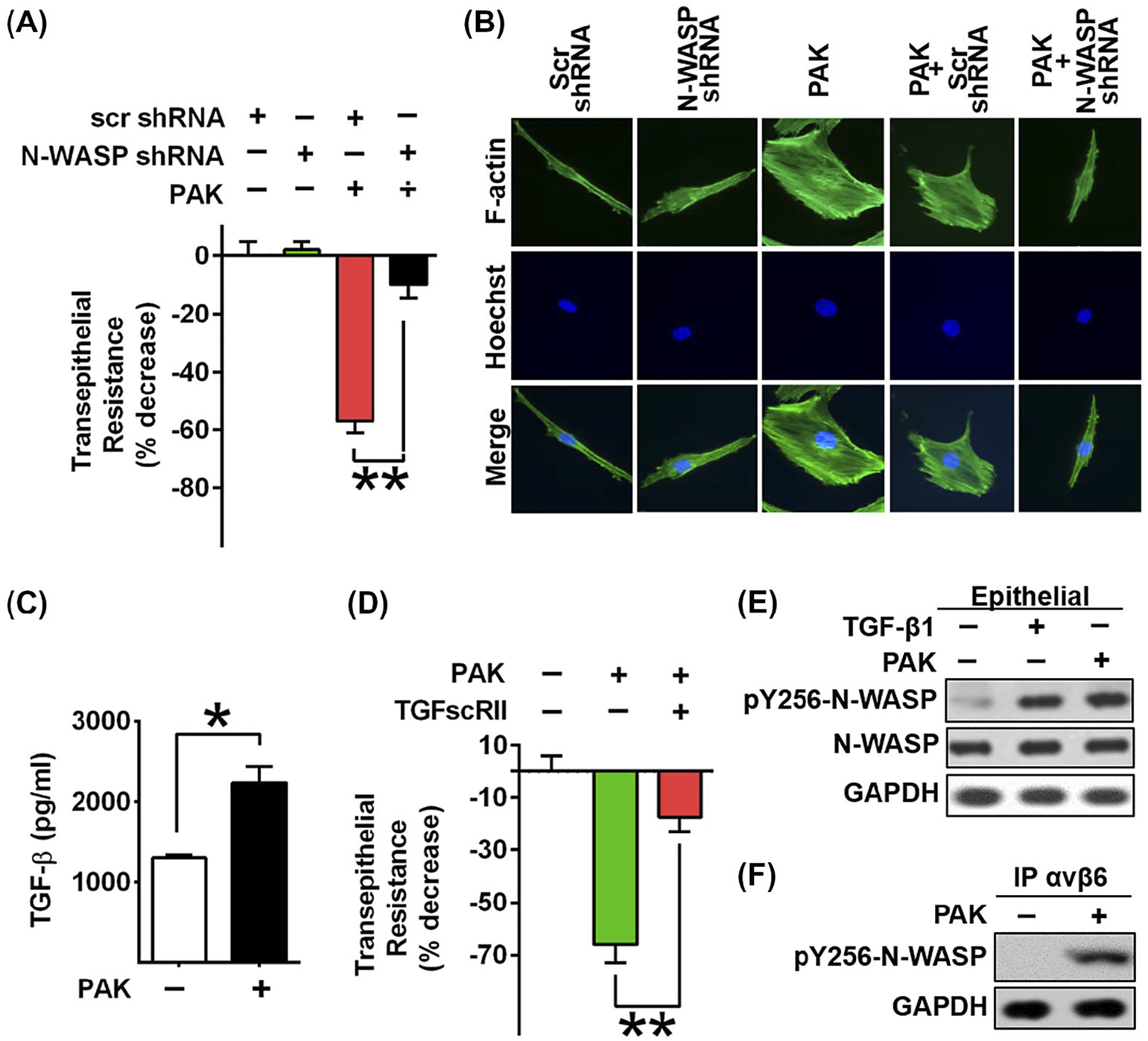
FIGURE 2.

N-WASP downregulation inhibited P aeruginosa induced permeability and cytoskeleton rearrangement in epithelial cells. A, Lung epithelial cells (L2) were challenged with P aeruginosa infection, followed by the measurement of transepithelial resistance with or without N-WASP downregulation. Resistance value was measured 24 h post P aeruginosa stimulation. Data are presented as the percentage of decreased resistance normalized to control (n = 6–8). B, Representative images of actin stress fiber formation in L2 cells transduced with lentivirus targeting N-WASP or scramble shRNA followed by P aeruginosa treatment. Actin stress fibers are immunostained with phalloidin (green) and nuclei with Hoechst (blue). The downregulation of N-WASP blocked P aeruginosa induced actin stress fiber formation. C, ELISA assays showed increased active TGF-β1 production upon P aeruginosa stimulation in L2 cells. D, Blockade of TGF-β1 signaling pathway by treatment of L2 cells with a soluble chimeric TGF-β type II receptor (10 ng/mL) inhibited P aeruginosa induced epithelial paracellular permeability in L2 monolayers (n = 6–8). E, Representative blot shows increased phosphorylation of N-WAPS Y256 in response to TGF-β1 and P aeruginosa treatment in L2 cells. F, Western blot of co-immunoprecipitation assays showed that P aeruginosa induced the association of integrin αvβ6 with active form of N-WASP. Data are presented as means ± SEM. *P < .01 and **P < .001
