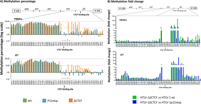
Fig 2. Deletion of the CTCF binding site of HTLV-1 (vCTCF-BS) results in expansion of DNA methylation in the pX region of the provirus.
DNA methylation of the HTLV-1 provirus is presented as the percentage of methylated CpG (A) or fold change (B) (Y-axis) at the indicated locations of the viral DNA (X-axis). The schematic diagram of HTLV-1 provirus indicates the regions examined by bisulfite treatment and DNA sequencing as described in the Materials and Methods. Upper panel: HTLV-1 immortalized bulk population of PBMCs; lower panel: HTLV-1 infected bulk population of JET cells. CTCF binding site: 7041-7052 as indicated by an arrow. * Lost CpG sites, ** New CpG site due to the introduced mutations in vCTCF-BS. Nine CpG sites (5649-6076), 19 CpG sites (6503-7014), 8 CpG sites (7398-7615) are not shown because of unsuccessful PCR amplification in these regions after bisulfite treatment.

