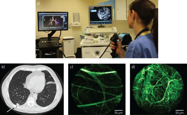
FIGURE 2.
a) The delivery of fibre-based optical endomicroscopy (OEM), in conjunction with navigational bronchoscopy, to access a distal solitary pulmonary nodule (SPN) in the clinical setting. Computed tomography (CT) and OEM images from an individual patient presenting with an SPN: b) CT demonstrating solid SPN; c) OEM image demonstrating surrounding healthy elastin structure; d) OEM image demonstrating distorted, abnormal elastin structure within the SPN (subsequently confirmed as benign on histopathological analysis) [35].

