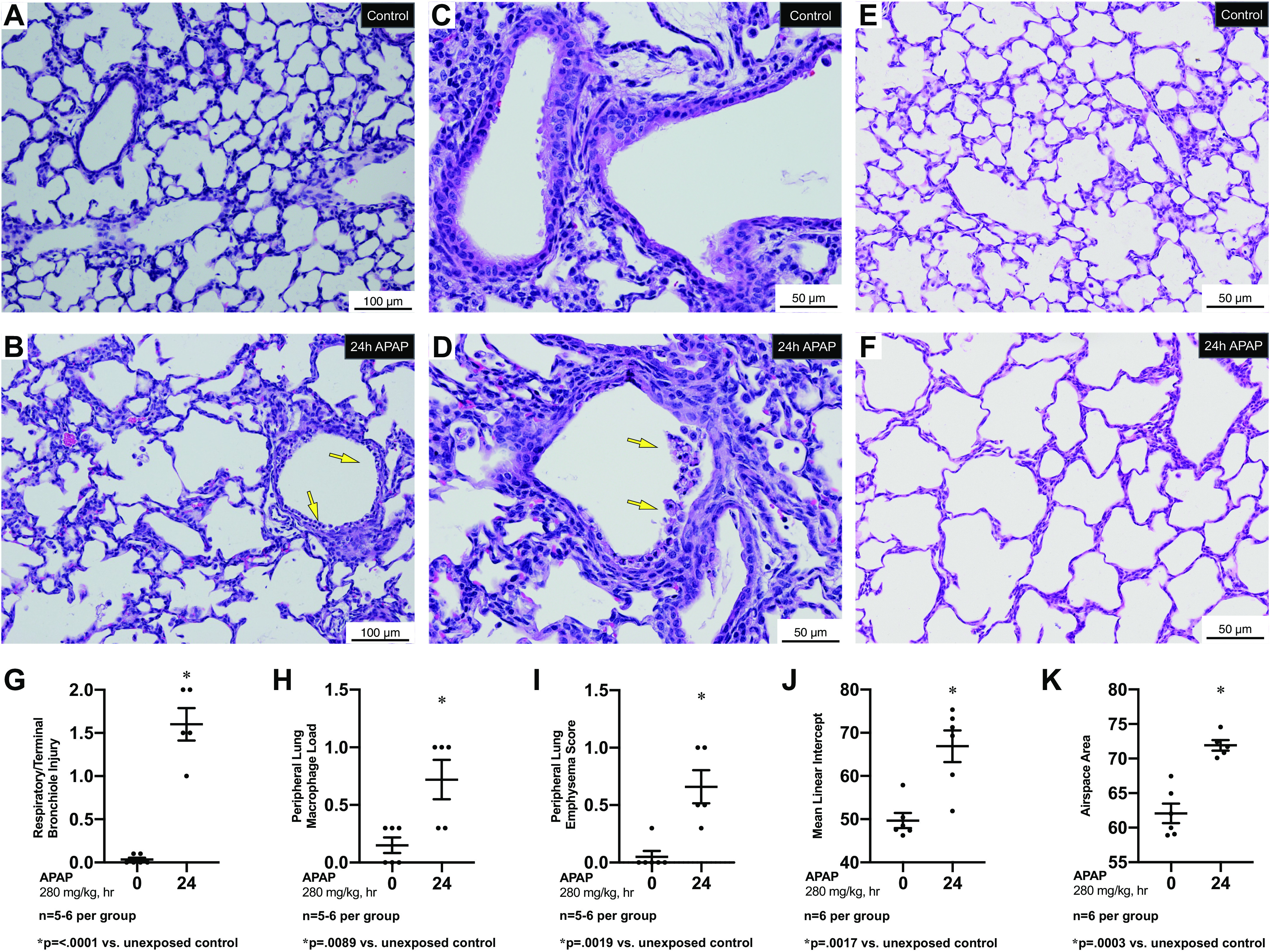
Figure 2.

Pulmonary response to APAP exposure (280 mg/kg, IP) at PN7. A–D: low (A, B) and high (C, D) magnification representative H&E-stained proximal sections from unexposed (A, C) and exposed (C, D) (APAP 280 mg/kg, IP; 24 h administered on PN7) C57BL/6 mice. Yellow arrows indicate cell death and sloughing of a portion of the surface epithelium lining a bronchiole. Internal scale bar 50 or 100 μm E and F: high magnification representative H&E-stained distal sections from unexposed (E) and exposed (F) (APAP 280 mg/kg, IP; 24 h administered on PN7) C57BL/6 mice. Internal scale bar 50 or 100 μm. G–I: blind histopathologic evaluation of H&E-stained pulmonary sections from unexposed and exposed (APAP 280 mg/kg, IP; 24 h administered on PN7) C57BL/6 mice scored for respiratory/terminal bronchiole epithelial injury (G), peripheral lung macrophage load (H), and peripheral lung emphysema score (I). Data are expressed as mean ± SE; J and K: morphometric assessment of H&E-stained pulmonary sections from unexposed and exposed (APAP 280 mg/kg, IP; 24 h administered on PN7) C57BL/6 mice for mean linear intercept (J) and airspace area (K). Data are expressed as mean ± SE. APAP, n-acetyl-p-aminophenol; H&E, hematoxylin-eosin; PN7, early alveolar stage of lung development.
