
FIGURE 2.
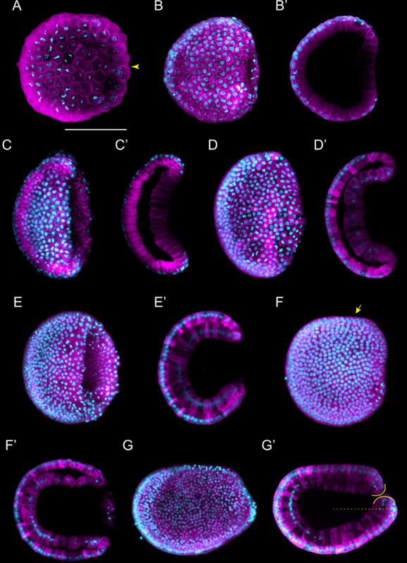
Branchiostoma lanceolatum gastrula stages. Embryos are stained with the lipophilic dye FM 4-64 (magenta) and with the DNA dye Hoechst (cyan). Animal pole and anterior are to the left and dorsal side is up. (A–G) Maximum projections of confocal z-stacks of entire embryos. (B′–G′) Single z-stacks highlighting the inner morphology of the developing gastrula. (A) G0 stage, (B,B′) G1 stage, (C,C′) G2 stage, (D,D′) G3 stage, (E,E′) G4 stage, (F,F′) G5 stage, (G,G′) G6 stage. In (A), the yellow arrowhead indicates the vegetal cells. In (F), the yellow arrow highlights the flattened side of the gastrula embryo. In (G′), the yellow lines delimit the upper and lower lips of the blastopore, and the dashed line indicates the midline of the embryo. Scale bar: 50 μm.