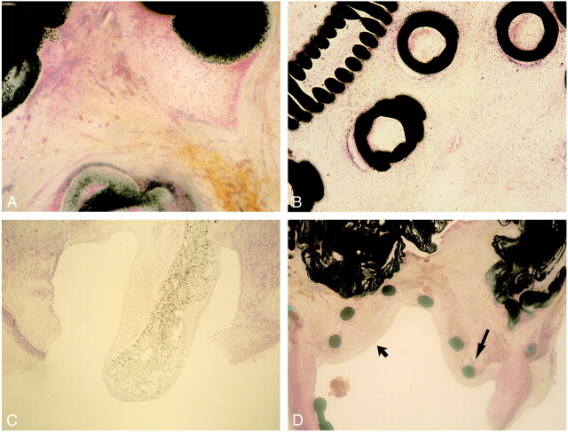
fig 6.
A, Light microscopic findings in region of aneurysmal dome 14 day after embolization with Onyx (hematoxylin-eosin, ×25). Dense connective tissue containing moderate number of mixed inflammatory cells is observed along with polymer surfaces. No angionecrosis is present.
B, Light microscopic findings in region of aneurysmal dome 14 days after embolization with GDCs (hematoxylin-eosin, ×25). Minimum to mild inflammatory reaction is observed along with coils.
C, Light microscopic findings of aneurysmal neck 14 days after embolization with Onyx. Note tonguelike migration of Onyx into the parent artery and complete coverage with neointima (hematoxylin-eosin, ×10).
D, Light microscopic findings of aneurysmal neck 14 days after embolization with Onyx and microstent. Note significant intimal hyperplasia (wide arrow) and stent deformity due to tissue reaction (thin arrow) (hematoxylin-eosin, ×5).

