
fig 6.
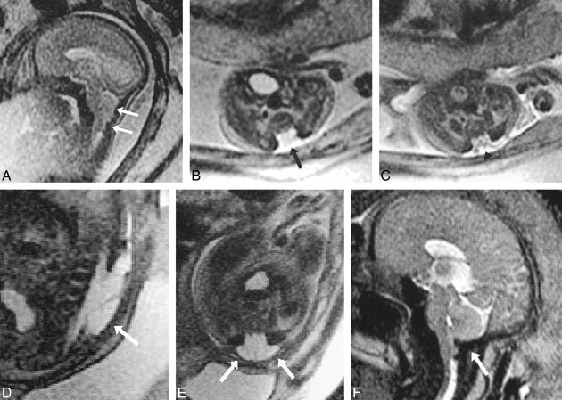
Patient 17: Chiari II malformation and myelomeningocele (23 weeks' gestation).
A, Sagittal ssFSE image (∞/96/0.5) shows the poorly formed posterior fossa floor and downward cerebellar herniation (arrows).
B and C, Axial ssFSE images at the level of the lumbosacral region show absent posterior elements (arrow, B) and exposed neural elements (myelomeningocele, arrowhead, C).
D and E, Sagittal (D) and axial (E) ssFSE images (∞/98/0.5) 13 days after in utero repair show hypointense dural patch (arrows) over defect.
F, Sagittal ssFSE image (∞/97/0.5) approximately 10 weeks after repair suggests improved development of the floor of the posterior fossa (suboccipital bone, arrow) and reduced hindbrain herniation.