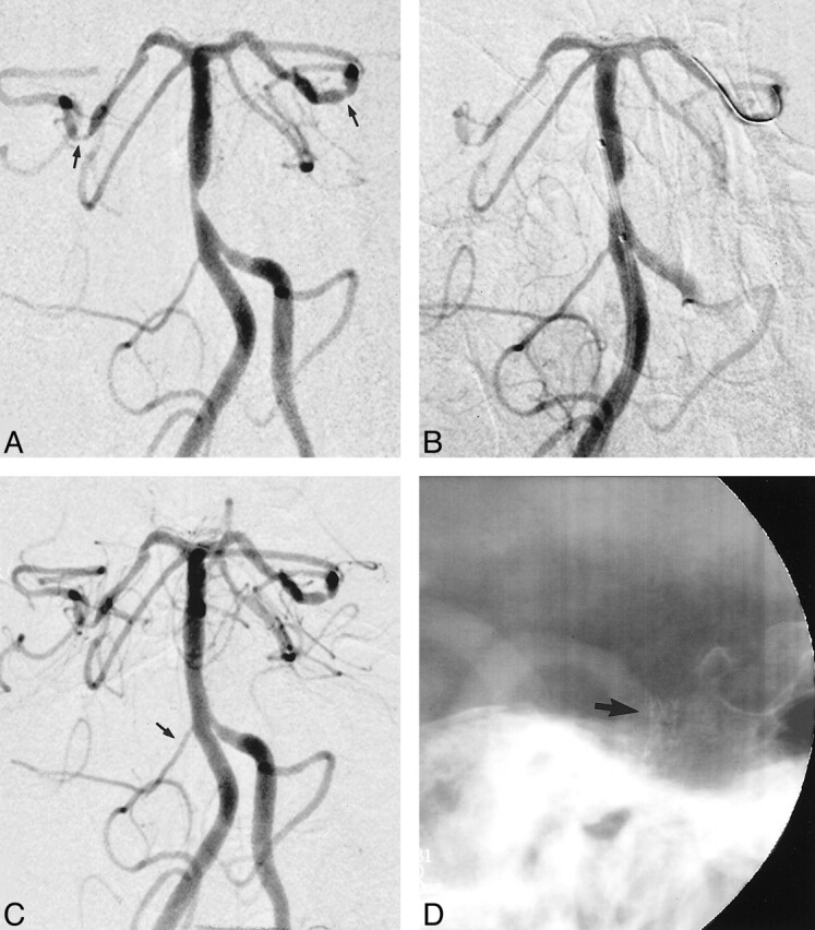
fig 1.

Anteroposterior oblique views of the basilar artery.
A, Eccentric high-grade (>75%) stenosis of the lower basilar artery above the origin of the right AICA. The left AICA arises from the left PICA. Note bilateral stenosis of the posterior cerebral arteries (arrows) and a stenosis involving the distal left vertebral artery at the vertebrobasilar junction.
B, Stent is positioned across the stenosis without preliminary balloon angioplasty.
C, After stent deployment, normal vessel lumen is restored. Although not obvious on this image, the inferior margin of the stent covers the right AICA origin; however, the vessel continues to opacify normally (arrow).
D, Nonsubtracted lateral oblique view of the posterior fossa shows the basilar artery metallic stent posterior to the clivus (arrow).
