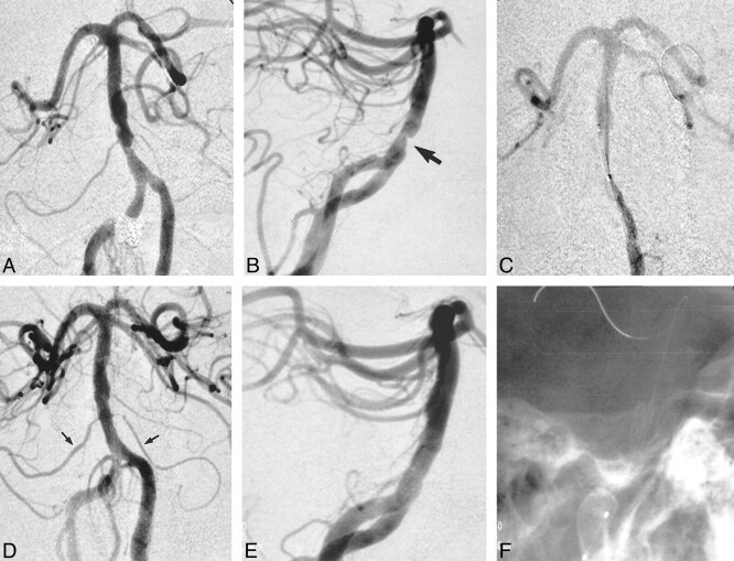
fig 2.
A and B, Anteroposterior (A) and lateral (B) views of the basilar artery show an eccentric high-grade (>70%) stenosis of the lower basilar artery projecting from the anterior wall (arrow, B). The AICAs originate immediately above the lesion.
C, Anteroposterior view of the basilar artery shows positioning of the stent across the lesion without preliminary balloon angioplasty. Note that positioning of the stent delivery catheter across the stenosis results in significant flow compromise, confirming the high-grade nature of this lesion.
D and E, Anteroposterior (D) and lateral (E) views of the basilar artery after endovascular stent deployment show restoration of normal vessel lumen. Although not obvious on these images, the superior margin of the stent covers the origins of both AICAs (arrows, D), which, however, continue to opacify normally.
F, Nonsubtracted lateral oblique view of the posterior fossa shows the basilar artery metallic stent. The tip of the stent delivery catheter has been retracted inferiorly with the 0.014-inch exchange guidewire still in place.

