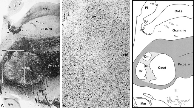
fig 2.

A, Sagittal section through the mesencephalon at the level of the red nucleus shows the subdivision of the parvicellular subnucleus into the pars oralis, pars dorsomedialis, and pars caudalis (Heidenhain stain; original magnification × 8).
B, Sagittal section through the mesencephalon at the level of the red nucleus shows the subdivision of the parvicellular subnucleus into the pars oralis, pars dorsomedialis, and pars caudalis (original magnification × 30). The corresponding area of the adjacent myelin-stained section is delineated by the square in A.
C, Drawing of red nucleus partitions according to A shows the medullary lamellae (ML) in the middle of the parvicellular subnucleus of the red nucleus.
Dm indicates pars dorsomedialis; Or, pars oralis; Caud, pars caudalis; Mm, mammillary body; Pe.ce.s, pedunculus cerebellaris superior; Pi, pineal body; III, rootlet of the oculomotor nerve; Col.s, colliculus superior; Gr. cn.me, griseum centrale mesencephli. Parts A and B were reproduced with permission from S. Karger, Basel, Switzerland.
