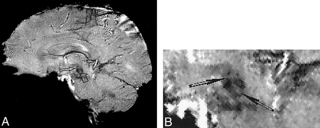
fig 4.

A, Typical oblique sagittal gradient-echo MR image (60/40/15) for one subject through the centers of the right red and dentate nuclei. Two lamellae (arrows) within the right red nucleus are clearly shown as cross-shaped structures. The signal intensity of the lamellae is relatively higher than that of the other parts of the red nucleus.
B, Enlarged view of the red nuclei. Arrows indicate the medullary lamellae.
