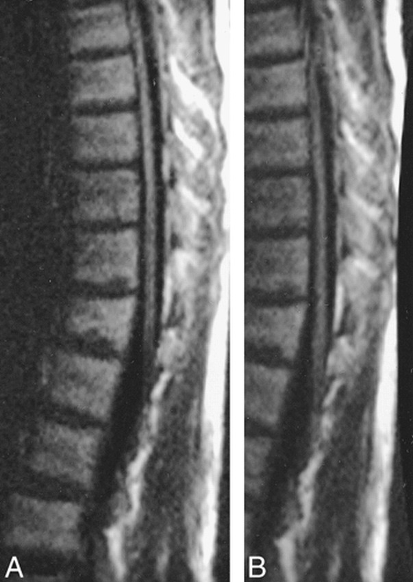
fig 1.

Comparison of LSDI images of the thoracic spine in a healthy adult volunteer shows comparable quality.
A, Half field of view (64 columns). Sagittal LSDI isotropic high b factor image (2912/76/1, 128 × 64 columns, 4-mm section thickness, b = 750 s/mm2 extrapolated to 1000 s/mm2, six directions) using a 30 × 15-cm field of view and an imaging time of 50 s per location.
B, Quarter field of view (32 columns). Sagittal LSDI isotropic high b factor image (1456/76/1, 128 × 32 columns, 4-mm section thickness, b = 750 s/mm2 extrapolated to 1000 s/mm2, six directions) using a 30 × 7.5-cm field of view and an imaging time of 25 s per location.
