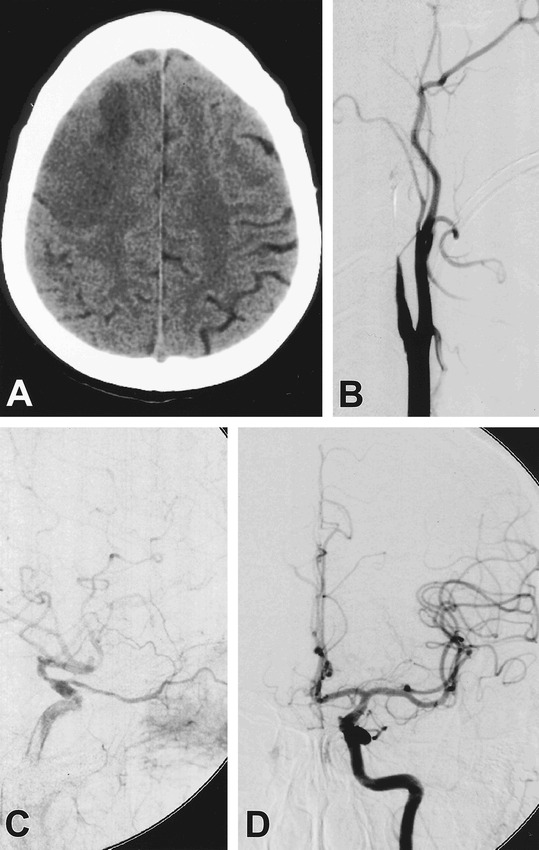
fig 2.

A 45-year-old woman (patient 1) noted to have developed left hemiparesis after diagnostic angiography at an outside hospital. Head CT scan shows evidence of a previous focal infarct as well as diffuse edema in the right posterior frontal lobe (A). Digital subtraction angiography of the right common carotid artery reveals tapering of the right internal carotid artery, to a complete occlusion, with appearance consistent of dissection (B). Injection of the right external carotid artery (C) shows retrograde collateral flow through the right ophthalmic artery, with filling of the cavernous segment of the right ICA. Injection of the left internal carotid artery shows no significant flow across the anterior communicating artery (D)
