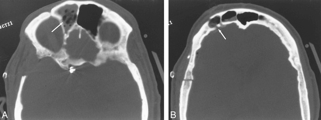
fig 2.

Sinus entry. A and B, These axial CT images filmed in bone windows are from a 56-year-old with an anterior communicating artery aneurysm who had extensive pneumatiztion of the frontal sinus and thick calvarium. Owing to frontal sinus entry during a right pterional craniotomy, the patient required frontal sinus mucosal exenteration with antibiotic-laden gelfoam (arrows) placed in the sinus. A pericranial graft over the entrance of the frontal sinus was also required
