fig 1.

Case 1.
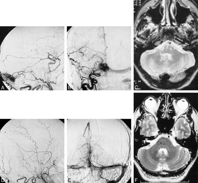
A, March 1996 angiogram. Injection of the right external carotid artery in lateral view shows the torcular dural AVF fed mainly by the occipital artery.
B, Same injection on anteroposterior view showing the dural AVF drainage through the left transverse sinus.
C, T2–weighted MR imaging at 2600/80/2 (TR/TE/excitations) performed during the same period showing torcular dilatation.
D, November 1999 angiogram. Lateral view of the right external carotid artery showing the disappearance of the fistula. E, November 1999 angiogram. Late phase of the left vertebral artery angiogram showing the normal patency of the torcular and the 2 transverse sinuses. F, November 1999 MR imaging (fast spin-echo T2-weighted [4900/106/2]) shows significant decrease in size of the torcular
