fig 5.
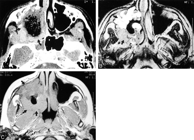
Images from the case of a 12-year-old male patient who complained of dental pain associated with nasal obstruction and progressive exophthalmos.
A, Initial contrast-enhanced CT scan shows a heterogeneous, necrotic mass involving the right maxillary sinus (arrows), invading the wall of the maxillary sinus, the right nasal fossa, and the right lateral pterygoid muscle.
B, T2-weighted MR image (3695/99/2) shows that the lesion is heterogeneous.
C, The lesion is isointense on the T1-weighted MR image (440/12/2). No recurrence had occurred 2 years after surgery, chemotherapy, and radiotherapy

