
fig 2.
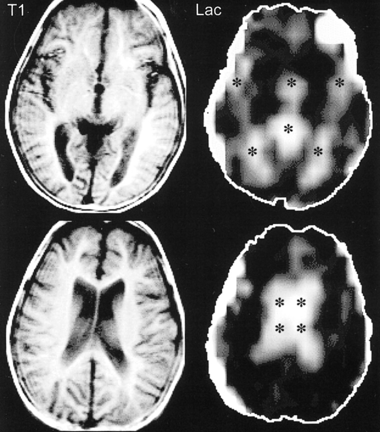
Patient 2. T1-weighted spin-echo MR images (300/13 [TR/TE]) at the level of the third ventricle (top row) and the lateral ventricles (bottom row) were unremarkable except for mild volume loss. Lac images showed high signal limited to the CSF spaces (*) (third and lateral ventricles, sylvian fissures, and cistern of the velum interpositum)