fig 1.
Continued.
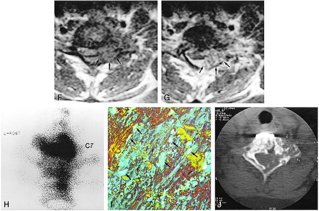
F and G, Axial T1-weighted MR images show the extradural mass encircling the anterior and lateral aspect of the dural sac (arrows, F). After contrast administration (G), the enhancing mass partially compresses the spinal cord (arrows).
H, Posterior pinhole bone scintigram of cervicothoracic junction obtained with 99mTc-MDP shows increased radioactivity in C7. Minimally increased radioactivity is also noted in T1 and T2 vertebral bodies.
I, Biopsy specimen viewed with polarized light is positive for Congo red stain, showing a characteristic green or apple-green birefringence (arrows) (original magnification ×80).
J, At 3-year follow-up, CT scan shows no growth of the residual mass in C7 vertebra (arrows). The metallic instrument for fixation is noted in the anterior aspect of C7.

