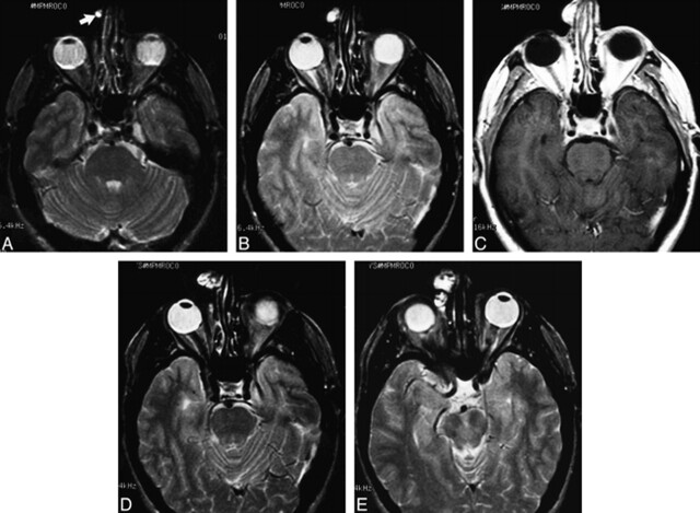
fig 1.
Case 1: MR findings in a 47-year-old man with clival chordoma.
A, Axial T2-weighted image (2500/80/1) shows a small, well-circumscribed, hyperintense nodule along the right side of the nose (arrow).
B, Axial T2-weighted image 6 months later shows interval enlargement of the mass involving the skin and subcutaneous tissues.
C, Contrast-enhanced axial T1-weighted image (600/13/2) shows intense enhancement of the mass.
D and E, Axial T2-weighted images 2 years later show progressive enlargement of the increasingly heterogeneous, lobulated, soft-tissue mass. At surgery, histopathologic findings were consistent with chordoma.

