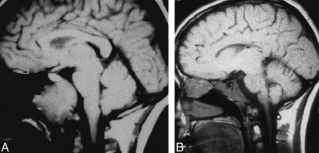
fig 3.
Case 3: MR findings in a 33-year-old woman with clival chordoma.
A, Sagittal T1-weighted image (600/11/3) obtained at initial presentation shows a large soft tissue mass centered on the clivus, consistent with known chordoma. The anterior ethmoidal region is unremarkable.
B, Sagittal T1-weighted image 2.5 years later shows abnormal signal in the central skull base, presumably related to postoperative/postradiation change, as it had been stable on several post-therapy imaging studies. In addition, a large soft-tissue mass has developed, centered on the anterior ethmoidal region. Recurrent chordoma was confirmed histopathologically.

