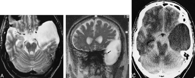
fig 5.
23-year-old man with a left-sided temporal arachnoid cyst, suffering from occasional headaches.
A, Transverse T2-weighted SE image (2000/80/1) shows a hyperintense left temporal arachnoid cyst.
B, Coronal PSIF image (20/25/1) depicts distinct signal reduction (arrow) arising from the basal cisterns, indicating probable communication.
C, CT cisternogram (3-hour delay) shows slight contrast enhancement within the cyst, confirming slow communication. The contrast enhancement was confirmed by measuring a clear increase of intracystic HU. The site of communication was not detectable on the CT cisternogram owing to proposed valve mechanism with slow communication.

