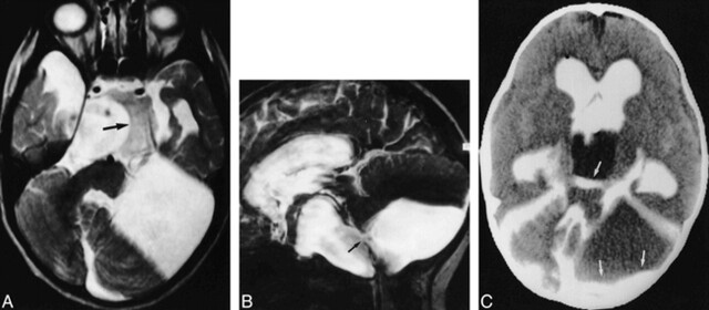
fig 6.
6-year-old girl with a system of temporal, supra- and parasellar, and paracerebellar arachnoid cysts.
A, Transverse T2-weighted SE image (2000/80/1) shows a right and small left temporal, suprasellar, and paracerebellar cysts. Note hamartoma of the tuber cinereum (arrow) anterior to the rotated brain stem.
B, Parasagittal PSIF image (20/25/1) shows some evidence of communication between the parasellar and paracerebellar cysts in the form of a continuous flow void (arrow).
C, CT cisternogram shows contrast enhancement with layering in the parasellar and paracerebellar cysts (arrows), providing further evidence of communication. Layering was caused by the patient's prolonged supine position.

