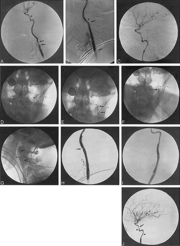
fig 2.

Patient 2. A–C, Angiography: ECA occlusion distal to small superior thyroidal and lingual branches, irregular narrowing in cervical ICA (arrows) and severe stenosis in vertical petrous ICA (A); Wallstent across carotid bifurcation (arrows) with ECA branches patent, improved CCA contour/caliber after deployment, but mild ICA spasm distal to stent (B); inflated AVE stent delivery balloon (arrows) shows reduced petrous ICA stenosis while stent is unattached to uninflated balloon (C). D–G, Fluoroscopy: AVE stent (arrowhead) with delivery balloon (short arrows) withdrawn over ACS guidewire into cervical ICA (D); 2-mm microsnare (small arrow) around ACS guidewire before AVE stent stabilization for balloon catheter (large arrows) removal (18-mm dime for measurement) (E); microsnare around AVE stent (large arrow) beside undeployed Palmaz-Schatz stent mounted on Courier balloon catheter (small arrows) (F); Palmaz-Schatz stent (arrows) deployed in distal ICA with crushed AVE stent (arrowhead) against distal cervical ICA and microsnare attached (G). H–J, Angiography: adequate ICA lumen adjacent to crushed, pinned AVE stent (arrow) after Palmaz stent deployment (H); AP view confirms adequate ICA lumen (I); minimal residual petrous ICA stenosis (arrow) and normal filling of MCA and ACA at procedure's end but irregular distal cervical and petrous ICA (arrowheads) shows residual spasm/dissection (J).
