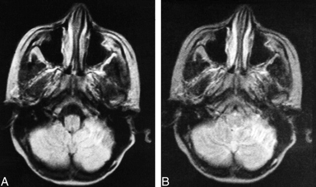
fig 2.
Images from the case of an 11-year-old female patient.
A, Proton density–weighted transverse spin-echo MR image (2200/50/2) shows a left cerebellar tuber with hypointense inner lines from calcification.
B, T2-weighted transverse spin-echo MR image (2200/100/2). There were 29 associated cerebral cortical tubers, three associated subependymal nodules, and six associated white matter nodules and lines.

