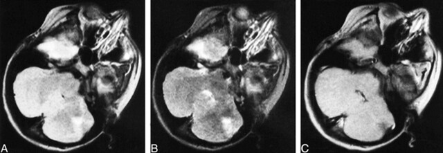
fig 3.
Images from the case of a 12-year-old male patient.
A, Proton density–weighted transverse spin-echo MR image (2200/50/2) shows a left cerebellar tuber with volume loss of the underlying parenchyma.
B, T2-weighted transverse spin-echo MR image (2200/100/2).
C, Contrast-enhanced T1-weighted transverse spin-echo MR image (350/30/2). There were 13 cerebral cortical tubers, eight subependymal nodules, and one white matter nodule.

