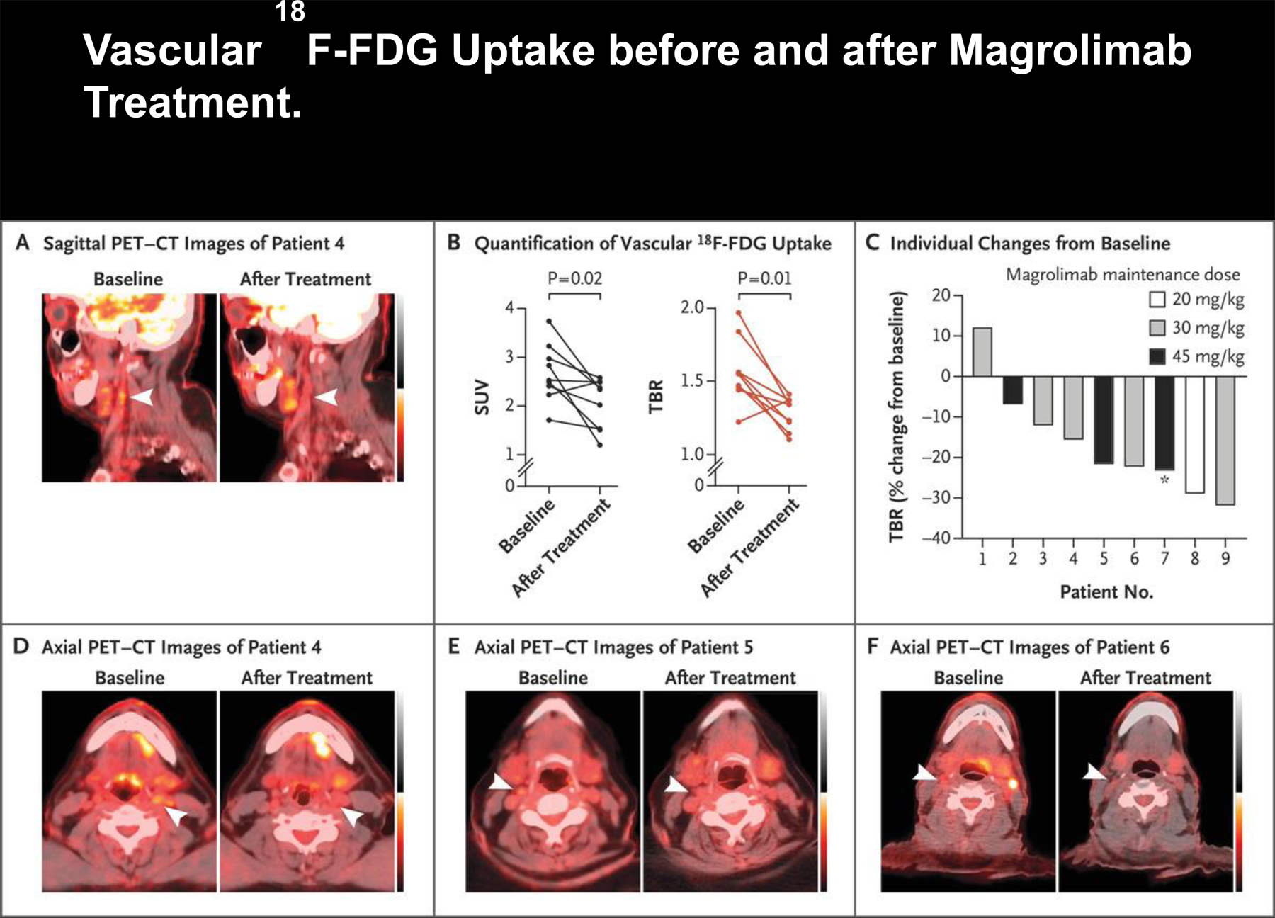
Figure 1. Vascular 18F-FDG Uptake before and after Magrolimab Treatment.

Panel A shows 18F-fluorodeoxyglucose (18F-FDG) positron-emission tomography and computed tomography (PET–CT) scans (sagittal view) of Patient 4, who had a reduction in vascular 18F-FDG uptake in the index carotid artery (arrowheads) after magrolimab treatment. Panel B shows the maximum standardized uptake values (SUVs) and target-to-background ratios (TBRs) in the most diseased segment of the index carotid artery in all nine patients at baseline and after magrolimab treatment. Panel C shows a waterfall plot of maximum TBR in all nine patients according to the maintenance dose received. Of note, for Patient 7 (asterisk), treatment was initiated at a maintenance dose of 45 mg per kilogram of body weight, but the dose was later changed to 30 mg per kilogram. Panels D, E, and F show 18F-FDG PET–CT scans (axial view) of Patients 4, 5, and 6, respectively. Arrowheads indicate the index vessel (carotid artery) at baseline and after magrolimab treatment. Data from each patient before and after treatment were compared and analyzed with a Wilcoxon matched-pairs signed-rank test (two-tailed).
