Figure 1.
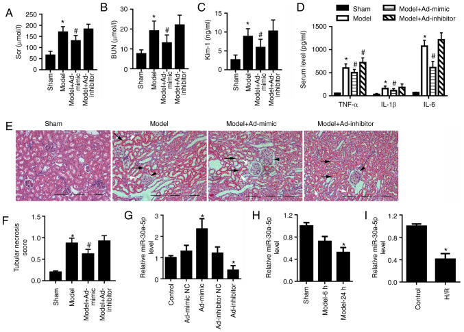
miR-30a-5p expression is decreased in in vivo and in vitro models of renal I/R injury. Mice were intraperitoneally injected with Ad-mimic or Ad-inhibitor 24 h before renal ischemia/reperfusion injury. ELISA analysis for serum levels of (A) SCr and (B) BUN, and (C) urine concentration of Kim-1 in mice (n=6). (D) ELISA analysis for serum levels of TNF-α, IL-1β and IL-6. (E) Renal histological changes in mice were observed by H&E staining; scale bar, 40 μm. Black arrows indicate the loss of epithelial cell nuclei; white arrows point to abscission of tubular epithelial cells; and black arrowheads indicate glomerular hypertrophy. (F) The histological tubular necrosis scores were evaluated. (G) miR-30a-5p expression levels in the cortex region of mouse kidneys following intraperitoneal injection with Ad-mimic or Ad-inhibitor were detected by RT-qPCR. (H) miR-30a-5p expression levels in the renal cortex region of mice was detected using RT-qPCR at 6 and 24 h post-reperfusion (n=6). (I) miR-30a-5p expression in H/R-exposed HK-2 cells was assessed by RT-qPCR (n=3). *P<0.05 vs. sham or control; #P<0.05 vs. model. Ad, adenovirus; H/R, hypoxia/reoxygenation; Kim-1, kidney injury molecule 1; miR, microRNA; RT-qPCR, reverse transcription-quantitative PCR; BUN, blood urea nitrogen; SCr, serum creatinine.

